Specialty Hub
Pulmonary Diseases
The diseases that fill RT shifts — from COPD, asthma, and ARDS to pneumonia, pulmonary embolism, and pneumothorax, plus the chronic and restrictive diseases (cystic fibrosis, bronchiectasis, interstitial lung disease, and pulmonary hypertension) — with management guides, references, and the comparison charts that keep the lookalikes straight.
Guides
Management of the obstructive, infectious, vascular, and chronic lung diseases.
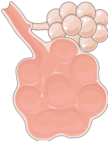
COPD Exacerbation Management
How RTs assess and manage an acute COPD exacerbation — controlled oxygen to 88–92%, combined SABA/SAMA bronchodilators, steroids, antibiotics, and NIV for hypercapnic respiratory acidosis.
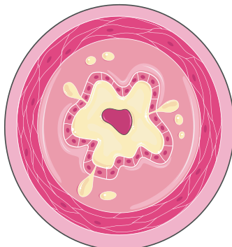
Asthma Exacerbation Management
Severity assessment and treatment of an acute asthma exacerbation — high-dose SABA, ipratropium, early steroids, oxygen, and the ominous signs of impending respiratory failure that a normal CO₂ can hide.
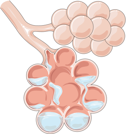
ARDS Basics
The Berlin definition, pathophysiology, and lung-protective ventilation of ARDS — 6 mL/kg of predicted body weight, plateau ≤ 30 cmH₂O, permissive hypercapnia, PEEP titration, and prone positioning.
Pneumonia: CAP, HAP, and VAP
Clinical guide to community-acquired, hospital-acquired, and ventilator-associated pneumonia: pathophysiology, severity scoring with CURB-65, assessment findings, and RT management priorities including oxygenation targets and VAP prevention.
Pulmonary Embolism
Pulmonary embolism obstructs the pulmonary arterial circulation, creating alveolar dead space, V/Q mismatch, and acute RV strain. Covers Virchow's triad, the PERC rule, capnography changes, risk stratification, and RT priorities for massive and submassive PE.
Pneumothorax and Chest Tube Management
Covers the full pneumothorax spectrum from primary spontaneous to tension, plus three-chamber drainage system management, air-leak troubleshooting, and ventilator barotrauma prevention for respiratory therapists.
Cystic Fibrosis
A clinical guide to cystic fibrosis covering CFTR pathophysiology, sweat chloride diagnosis, daily airway clearance techniques, the correct inhaled therapy sequence, and disease-modifying CFTR modulators for respiratory therapists.
Bronchiectasis
A clinical guide to non-CF bronchiectasis covering the vicious cycle of infection and inflammation, HRCT diagnosis with the signet-ring sign, daily airway clearance techniques, mucoactive therapy, and antibiotic strategies for stable disease and exacerbations.
Interstitial Lung Disease and Pulmonary Fibrosis
Interstitial lung disease inflames and scars the lung interstitium, producing a restrictive defect with impaired diffusion. Covers IPF, the UIP pattern, oxygen therapy, antifibrotic awareness, and PFT interpretation for respiratory therapists.
Pulmonary Hypertension
Pulmonary hypertension is elevated pressure in the pulmonary circulation that strains and ultimately fails the right ventricle. Covers the WHO five-group classification, hemodynamic definition, the diagnostic role of right heart catheterization, and why group-specific therapy matters.
Interactive Practice
Practice Tools
Interpret the gases and quantify the oxygenation behind every exacerbation.
References
Drug therapy and severity classifications at a glance.
COPD Pharmacologic Management
COPD drug therapy — maintenance bronchodilators and inhaled corticosteroids by class, GOLD group-based initial therapy, and the pharmacology of an acute exacerbation, with bedside RT notes.
Asthma Stepwise Management
GINA stepwise asthma therapy — the Track 1 (ICS-formoterol reliever) and Track 2 (SABA reliever) pathways across steps 1–5, controller and reliever roles, and exacerbation pharmacology.
Pulmonary Disease Severity Classifications
The severity systems RTs must recognize — COPD GOLD spirometric grades, asthma severity and control, the ARDS Berlin definition, and CURB-65 for pneumonia — in one scannable reference.
Quick Charts
The comparisons that separate the lookalikes.
COPD vs Asthma Chart
Side-by-side comparison of COPD and asthma — onset, reversibility, triggers, inflammation, disease course, and acute treatment overlap — with the ABG red flags that separate them.
Obstructive vs Restrictive Patterns Chart
Side-by-side comparison of obstructive and restrictive PFT patterns — FEV1, FVC, the FEV1/FVC ratio, TLC, RV, DLCO, and flow-volume loop — with the hallmark that defines each and the diseases behind them.
ARDS vs Cardiogenic Pulmonary Edema Chart
Side-by-side comparison of ARDS and cardiogenic pulmonary edema — mechanism, onset, chest film, BNP, echo, edema fluid, P/F ratio, response to diuresis, and management.
Suggested Learning Path
A path across the obstructive, infectious, vascular, pleural, and restrictive diseases.
Related Specialties
Pulmonary disease management connects directly to these areas.